
The unique physical and chemical properties of nanomaterials stem from size effects and ultrastructures, so it is important to observe the surface morphology of nanomaterials. Some detection methods and characterization methods for directly detecting the particle size of drugs can be used to observe the appearance of particles, including Scanning electron microscope (SEM), transmission electron microscope (TEM), atomic force microscope (AFM), and scanning tunneling microscope (STM). Non-intuitive methods such as nuclear magnetic resonance and differential thermal analysis can also be used to study the appearance of nanoparticles.
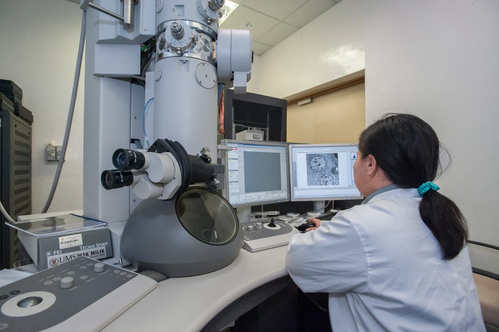
1. Scanning electron microscope
Scanning electron microscopes have a wide range of magnifications, ranging from several times to hundreds of thousands of times, covering the magnification range of optical magnifiers to transmission electron microscopes, and the resolution is very high. The scanning electron microscope has a large focal depth of 300 times than that of an optical microscope. For complex and rough sample surfaces, clear and focused images can still be obtained. SEM sample preparation is relatively simple, and material samples need only be simply cleaned and coated to observe, and the sample size requirements are very low.
2. Transmission electron microscope
Transmission electron microscopy can be used to observe the morphology, dispersion of nanoparticles, and measure and evaluate particle size. Atomic-level appearance can be obtained by TEM, and its resolution is about 1 nm. The combination of line-scan mode transmission electron microscopy and electron energy loss spectrometer (EELS) can be used to analyze nanoscale multilayer structures. TEM samples are required to be nanometer-thick films that can be penetrated by the electron beam, and are preferably dispersed without agglomeration.
3. Atomic force microscopy
Atomic force microscopes belong to the scanning probe microscope (SPM) series. Compared with SEM and TEM, AFM is a high-resolution electron microscope (High resolurion EM, HREM). The advantages of AFM include: 1) less requirements for working environment and sample preparation than electron microscope, and it can detect the shape, size and mechanical properties of conductors, semiconductors, insulators and biological samples in the atmosphere, high vacuum, liquid and other environments; 2) extremely high resolution. The horizontal resolution is less than 0.1nm,and the vertical resolution is less than 0.01nm; 3) physical and mechanical properties of materials can be measured at the nanometer scale, such as electrical conductivity, stagnation, friction, and lubrication.
4. Ray diffraction
Ray diffraction includes powder X-ray diffraction (XRD), small angle X-ray scattering (SAXS), small angle neutron scattering (SANS), and electron diffraction (ED).
XRD is an effective means to identify the crystal phase of a substance. XRD is indispensable for the characterization of nanomaterials based on the position of a characteristic peak. XRD is also used for crystal structure analysis. According to the powder diffraction pattern, the position of the atoms in the unit cell, the parameters of the unit cell, and the number of atoms in the unit cell can be determined. High-resolution powder X-ray diffraction (HRXRD) provides more detailed structural information, and can obtain data and information on the fine structure of nanomaterials such as the elemental composition ratio, size, ion spacing, and bond length of related substances in single crystal cells.
SAXS can effectively detect the fractal structure of nanoparticle agglomeration, determine its fractal dimension, the average radius of agglomerates and primary particles, and is suitable for characterizing the structural characteristics of amorphous materials at relatively low resolution. The advantages of SAXS include a wide sample range, being non-destructive and suitable for dry and wet samples, no special sample preparation required, and the ability to characterize samples that cannot be measured by TEM. Sensitive detection can directly measure bulk materials, and has a better statistical average of particles.
5. Thermal analysis
Thermal analysis includes differential thermal analysis (DTA), differential scanning calorimetry (DSC), and thermal gravimetry (TG). The three methods are often combined and also combined with XRD, NMR, etc. They can be used to characterize: 1) the presence or absence, content, and thermal weight loss temperature of surface-bonding or non-bonding organic groups or other substances; 2) the relationship between the strength of the surface adsorption capacity (the amount of adsorbed substances) and the particle size; 3) particle size change during heating; 4) phase transition and crystallization during heating. As a useful supplement to XRD, this type of method is often used to determine the degree of heterogeneous coexistence in nanoparticles and the degree of interaction between various components in nanoparticles. DSC research on the physical state of triamcinolone acetate nanostructured lipid carrier (NLC) found that the crystallinity of solid lipids in the NLC system was significantly reduced, and drugs may exist in the amorphous or molecular state in the NLC system.
6. Nuclear magnetic resonance
Nuclear magnetic resonance (NMR), as an aid to the study of the structure of nanoparticles, can reflect the dispersion and compatibility of nanoemulsions in water, as well as the state of each component molecule in the colloidal system. After studying the microstructure of triptolide alcohol new solid lipid nanoparticles by NMR, it was found that the new solid lipid nanoparticles have good water dispersibility and compatibility. The sharpness of the signal between the nanoparticles is between the solid and liquid, indicating that the degree of freedom of molecular movement is between the liquid and the solid. The nanoparticles are composed of a combination of liquid oil and solid lipids.
Reference:
1. Panyam J, Labhasetwar V. Biodegradable nanoparticles for drug and gene delivery to cells and tissue. Adv Drug De-livery Rev, 2012, 64: S61-S71.