
Raman spectrum is a type of scattering spectrum. Raman spectroscopy is based on the Raman scattering effect found by Indian scientist CV Raman, and analyzes the scattering spectrum with a frequency different from the incident light to obtain information on molecular vibration and rotation. This analytical method is applied to the study of molecular structure.
Japanese scientists have developed a new Raman spectroscopy method that enables researchers to analyze the chemical composition and structure of metal particles with a diameter of only 0.5-2 nanometers. This latest breakthrough is expected to enable scientists to develop new micromaterials, which are widely used in electronics, biomedicine, chemistry, and other fields.
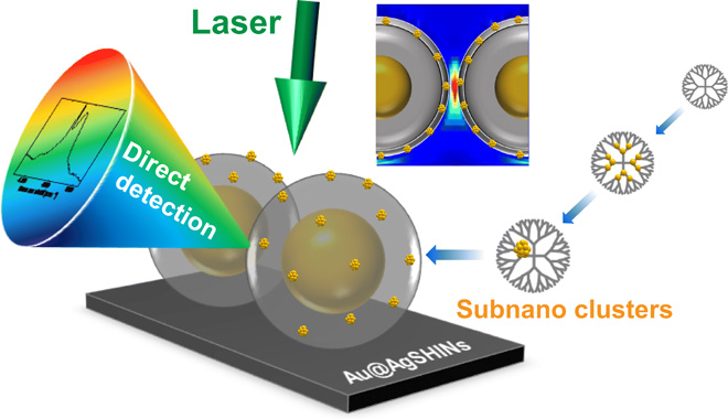
Metal nanoparticles have a wide range of potential applications and are becoming a hot material in modern research. Researchers have been able to develop metal nanocrystals with a diameter of only 0.5-2 nanometers (one nanometer is one billionth of a meter). These small particles are called “subnano clusters” (SNCs) and have very unique characteristics varying with the number of constituting atoms (atomicity). For example, it can act as an excellent catalyst in electrochemical reactions; it also exhibits a special quantum size effect. However, the existing analysis methods are not capable of SNCs detection research. For example, although traditional Raman spectroscopy and its variants have been used in many fields, it is not sufficient to detect SNCs due to its low sensitivity. In view of this, the Tokyo Institute of Technology research team proposed a new method to enhance the performance of Raman spectrometry and make it capable of SNCs analysis.
In the research, the Japanese team worked to improve the performance of specific Raman spectroscopy, surface-enhanced Raman spectroscopy. They said that adding gold/silver nanoparticles encapsulated in a thin shell of inert silica to the sample can amplify the light signal of the sample, thereby increasing the sensitivity of the technology. Therefore, they first theoretically determined the optimal size and composition of gold/silver and found that a 100 nm silver optical amplifier can greatly amplify the signal of SNCs adhered to a porous silica shell.
“This spectroscopic technique selectively generates Raman signals of substances that are situated in close proximity to the surfaces of the shell-isolated nanoparticles”, explains Professor Akiyoshi Kuzume, one of the study leaders. To verify these findings, they measured the Raman spectra of tin oxide SNCs to see if they could find an explanation in their structure or chemical composition to explain that they have unexplained high catalytic activity in certain chemical reactions. By comparing the results of Raman measurements with structural simulation and theoretical analysis, they discovered new insights into the structure of tin oxide SNCs, explaining the origin of the specific catalytic activity of tin oxide SNCs with atomic dependence The methodology used in this study will have a major impact on the development of better analytical techniques and sub-nanometer science. Professor Kimihisa Yamamoto concluded, “The high-sensitivity characterization methodology elaborated here can provide a comprehensive understanding of the chemical and structural natures of subnanomaterials, which facilitate the rational design of subnanomaterials on the atomic scale for practical applications, such as in catalysts, biosensors, and electronics.”
Reference:
Kuzume, A., Ozawa, M., Tang, Y., Yamada, Y., Haruta, N., & Yamamoto, K. (2019). Ultrahigh sensitive Raman spectroscopy for subnanoscience: Direct observation of tin oxide clusters. Science Advances, 5(12), eaax6455.