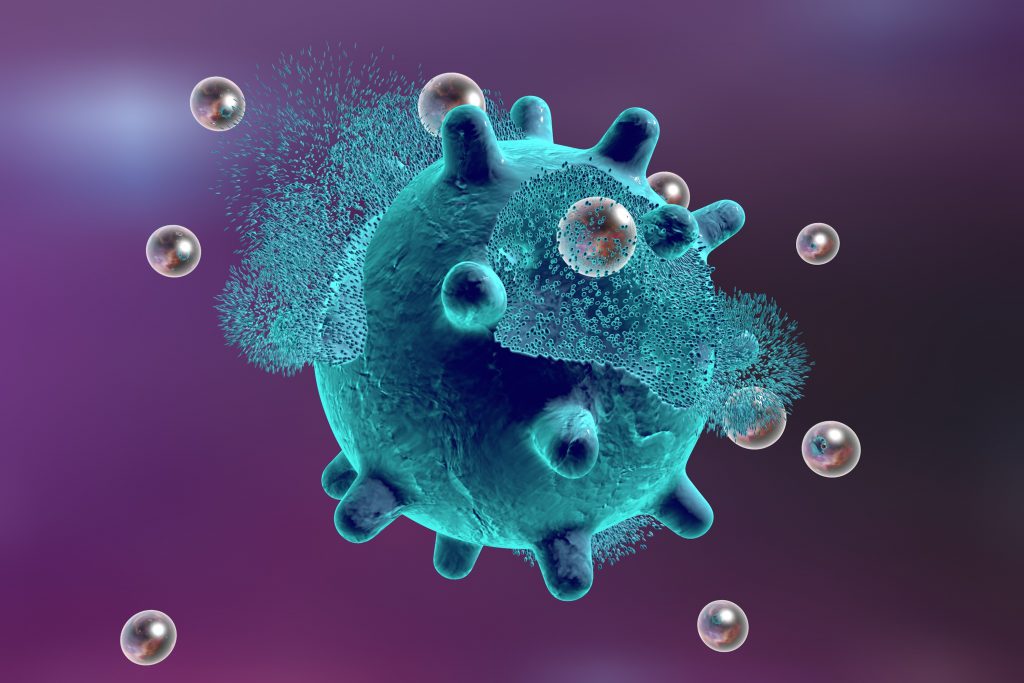
Metal nanoparticles have been widely used in broad applications in various fields. Recently, silver nanoparticles (AgNPs) attract a lot of attention due to their superior physical, chemical, and biological properties as well as the low-cost and abundance of raw materials. AgNPs have shown their great potential in anti-bacterial, anti-cancer therapeutics, diagnostics, optoelectronics, water treatment, and other clinical applications. There are studies shown that the physical, optical, and catalytic properties of AgNPs can be strongly affected by their size, distribution, morphological shape, and surface properties. The size of AgNPs can be tailored to specific applications. For example, for drug delivery, AgNPs are usually synthesized greater than 100 nm so that a proper quantity of drug can be delivered. The morphological shape of AgNPs could be in various shapes such as rod, triangle, round, octahedral, and polyhedral. As for the surface properties of AgNPs, they are generally modified by diverse synthetic methods, reducing agents and stabilizers. These exceptional properties of AgNPs have enabled their use in the fields of nanomedicine, pharmacy, biosensing, and biomedical engineering. In this article, we attempt to present you a brief introduction of silver nanoparticles about their synthesis and characterization.
Synthesis
- Physical method
The evaporation–condensation approach and the laser ablation technique are usually used for the physical synthesis of AgNPs. These two methods can fabricate a large quantity of AgNPs which are highly pure and chemicals releasing toxic substances or jeopardizing human health and environment will not be used during synthesis. In addition to these advantages, there are also several challenges: 1) agglomeration would occur due to the non-addition of capping agents; 2) greater power, relatively longer duration of synthesis and complex equipment are required when fabricating, leading to an increased operating cost.
- Chemical method
Metallic NPs can be synthesized by reducing their metal salts in an aqueous (or organic) solution to be a colloidal dispersion. Using this strategy, many metallic salts are already applied to synthesize corresponding metal NPs including gold, silver, iron, zinc oxide, copper, palladium, and platinum. For the chemical synthesis of AgNPs, they are usually fabricated via the Brust-Schiffrin or the Turkevich method. Moreover, with the presence of reducing and capping agents, the characteristics of AgNPs such as size distribution, shape, and dispersion rate can be easily changed or modified. In order to avoid aggregation, stabilizing agents will also be used. During the synthesis, several factors should be considered for a safer and more effective fabrication, including the choice of solvent medium, the use of an environment-friendly reducing agent and the selection of relatively non-toxic substances.
- Green method
The green method refers to the metal NP synthesis method using biological entities (microorganisms and plant extracts). Green synthesis is a promising alternative to physical and chemical methods because no toxic chemicals agents are used and no hazardous byproducts will be produced and those capping agents for the stabilization of AgNPs are all natural. Microorganisms such as bacteria and fungi have been utilized for the remediation of toxic materials by reducing metal ions. In these bacteria, their intracellular components function as the reducing and stabilizing agents when synthesizing AgNPs.
Characterization
The characterization of AgNPs is to determine the phase purity, shape, size, morphology, electronic transition plasmonic character, atomic environment, surface charge, etc. Several advanced analytical techniques are involved including electron microscopic techniques (atomic force microscopy (AFM), electron energy loss spectroscopy (EELS), surface enhanced Raman scattering (SERS), scanning electron microscopy (SEM) and transmission electron microscopy (TEM) and their corresponding energy-dispersive X-ray spectroscopy (EDX), and selected area electron diffraction (SAED for crystallinity). Surface morphology, size, and overall shape can be determined by SEM-/TEM-/EELS-supported EDX. There are many other techniques applied to determine the size distribution, dispersibility, average particle diameter of AgNPs:
- Fluorescence correlation spectroscopy (FCS): diffusion coefficients, hydrodynamic radii, average concentrations, and kinetic chemical reaction;
- X-ray diffraction (XRD): phase purity with crystal parameters and particle size;
- UV-Vis spectroscopy: band gap, particle size electronic interaction;
- X-ray photon spectroscopy (XPS): surface environment of elemental arrangement;
- Raman spectroscopy: submicron spatial resolution average size and size distribution;
- Nuclear magnetic resonance (NMR): structure, compositions, diffusivity of nanomaterials and dynamic interaction of species under investigation;
- Small-angle X-ray scattering (SAXS): size distribution, shape, orientation, and structure of a variety of polymers and nanomaterials.
References:
1. Chouhan, N. (2018). Silver Nanoparticles: Synthesis, Characterization and Applications. Silver Nanoparticles: Fabrication, Characterization and Applications, 21.
2. Lee, S. H., & Jun, B. H. (2019). Silver Nanoparticles: Synthesis and application for nanomedicine. International journal of molecular sciences, 20(4), 865.
3. Siddiqi, K. S., Husen, A., & Rao, R. A. (2018). A review on biosynthesis of silver nanoparticles and their biocidal properties. Journal of nanobiotechnology, 16(1), 14.
