
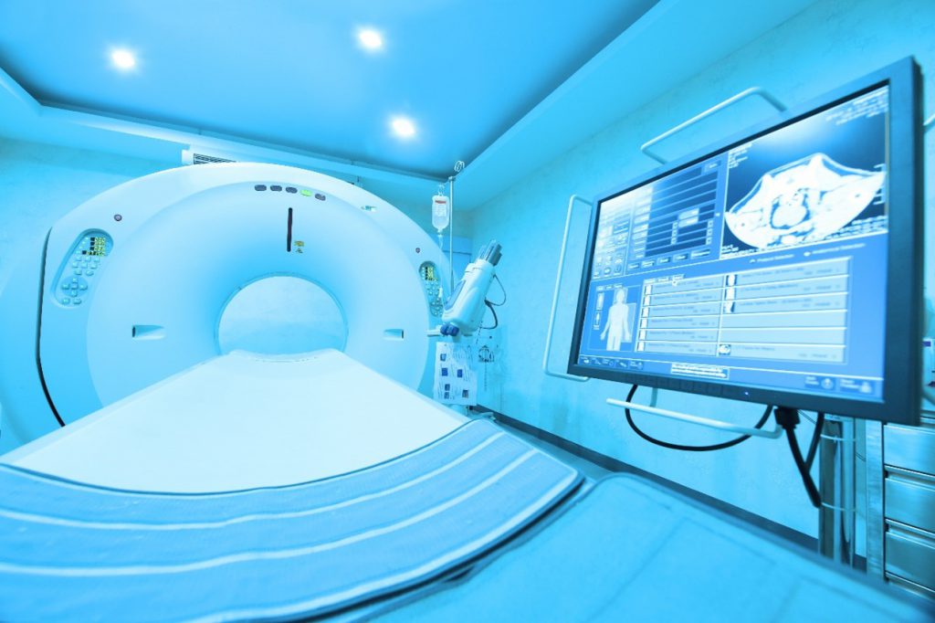
Magnetic resonance imaging (MRI) is a scan that uses a strong magnetic field and radio waves to generate detailed images of the inside of the body. MRI can be used to detect the brain tumors, traumatic brain injury, dysplasia, multiple sclerosis, stroke, dementia, infection, and causes of headaches. It is a noninvasive method that allows for fast and reproducible measurements of adipose tissue content in neonates with low intra- and intercoefficients of variability.
MRI is based on nuclear magnetic resonance (NMR), which was first documented in 1939 in a molecular beam by Isidor Rabi, who received the Nobel Prize in Physics in 1944. In 1946, techniques were developed independently by Felix Bloch and Edward Purcell that extended NMR to liquids and solids. Bloch and Purcell shared the Nobel Prize in Physics in 1952 for other important contributions to methods in magnetic resonance. It was not until 1973 that Paul Lauterbur devised a technique to create the first 2-D image from NMR signals. This is now known as MRI.
- Which is better a CT scan or MRI?
Both MRI and computed tomography (CT) scans can view the internal structure of the body. However, CT scans are faster and can provide pictures of tissues, organs, and bone structures. MRI is very good at capturing images and helping doctors determine whether there are abnormal tissues in the body. MRI is more detailed in its images, and crucially, it differs from CT by producing excellent soft tissue contrast without harmful ionizing radiation.
- What are contrast agents for MRI?
MRI is a non-invasive clinical imaging modality and has become widely used in the diagnosis and/or staging of human diseases around the world. Some MRI examinations include the use of contrast agents. The categorizations of currently available contrast agents have been described according to their effect on the image, magnetic behavior and biodistribution in the body, respectively. In this field, superparamagnetic iron oxide particles and soluble paramagnetic metal chelates are two main classes of contrast agents for MRI.
Gadolinium is the most famous MRI contrast agent. And iron oxide nanoparticles are the most prominent probes in MRI because of their tunable properties, such as magnetic properties, size, and easy binding to biomolecules. Iron oxide nanoparticles can also be combined with other imaging motifs such as fluorescent molecules and radioisotopes to create a multi-modal imaging system.
With years of experience in the development of diagnostic and imaging contrast agents, CD Bioparticles’ scientists can fine-tune physical parameters such as the size, shape, crystallinity, and magnetic properties of nanoparticles to improve their performance in MRI. In addition, Creative Diagnostics provides multi-modal imaging probes by combining iron oxide nanoparticles with fluorescent probes, PET probes, and ultrasound probes to improve the quality of diagnosis.
About the author
CD Bioparticles offers target-specific MRI contrast agents based on both gadolinium-chelate and iron oxide nanoparticles. Scientists in CD Bioparticles are proficient in design targeted nanoparticles as contrast agents for preclinical MRI. In addition, multimodal imaging contrast probes for MRI-optical and MRI-PET can be constructed to fit for your unique requirement.
References
Yan, G. P., Robinson, L., & Hogg, P. (2007). Magnetic resonance imaging contrast agents: overview and perspectives. Radiography, 13, e5-e19.
Narayan, R. (2018). Encyclopedia of biomedical engineering. Elsevier.