Although magnetic nanoparticles have been increasingly used in cell imaging and tissue bioengineering, the changes that have taken place within stem cells in the long-term have not been revealed. A recent study from CNRS, the Sorbonne University, and universities Paris Diderot and Paris 13, has shown that these nanoparticles undergo significant degradation inside the stem cells, and in some cases, the cells “remagnetize”. The results were published on PNAS, which may explain the existence of “natural” magnetism in human cells and help to develop new tools for nanomedicine, due to this magnetism generated by the cells themselves.
Magnetic nanoparticles are at the core of today’s nanomedicine: they act as imaging diagnosis agents, thermal anticancer agents, drug targeting agents and tissue engineering agents. After completing the therapeutic task, their fate in cells has not been well understood.
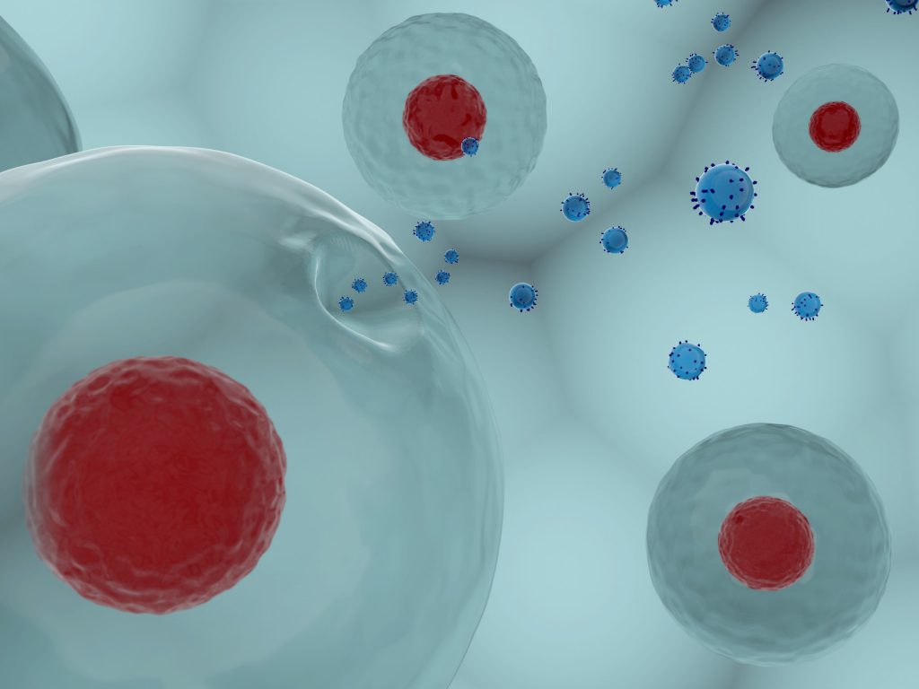
To track the journey of these nanoparticles through the cells, scientists from CNRS/University of Diderot and the INSERM/University of Paris Diderot/University of Paris 13, collaborated with researchers at Sorbonne University and together conducted the intensive research. Previously, researchers have developed a primitive approach to nanomagnetism in biological systems: first, they incorporated magnetic nanoparticles into human stem cells in vitro. Then researchers let them differentiate and develop for a month, observing them in the intracellular environment for a long time, and monitoring their transformation.
By tracking the “magnetic fingerprints” of these nanoparticles in cells, researchers have demonstrated that they are first destroyed (decreased cell magnetization) and release iron into the intracellular environment. Next, this “free” iron is stored in non-magnetic form in ferritin, which is responsible for storing iron or served as the basis for the biosynthesis of new magnetic nanoparticles in the cell.
This phenomenon is known to occur in some bacteria, but this biosynthesis has never been shown in mammalian cells. This could explain the magnetic crystals are present in the human body, which are observed in cells of different organs, especially the brain. What’s more, this magnetic iron storage could also be a way for cells to “detoxify” against the excess iron for a long time. From a nanomedicine perspective, this biosynthesis opens up a new avenue for the possibility of purely biological magnetic marking in cells. CD Bioparticles provides highly uniform nanoparticles for biology and medicine research uses, including colloidal gold nanoparticles, silver nanoparticles, silica nanoparticles, titania nanoparticles, inorganic fluorescent nanoparticles (quantum dots and upconversion nanocrystals), and biodegradable polymer nanoparticles. These nanoparticles are manufactured with different shapes and sizes, and the particle surface can be coated, functionalized or conjugated with biomolecules. Scientists focus on the fields of immunoassay, bioseparation, medical imaging and diagnosis, as well as drug delivery and cancer therapy can find more available nanoparticles at CD Bioparticles.
Reference:
Van de Walle, A., Sangnier, A. P., Abou-Hassan, A., Curcio, A., Hemadi, M., Menguy, N., … & Wilhelm, C. (2019). Biosynthesis of magnetic nanoparticles from nano-degradation products revealed in human stem cells. Proceedings of the National Academy of Sciences, 116(10), 4044-4053.